Lupus erythematosus

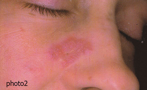
Discoid lupus erythematosus
1. FACE
The eruption consists of erythematous patches covered with an adherent hyperkeratotic layer, predominantly at the hair follicles. It resolves into cicatricial atrophy (photo 1). A rare form is lupus erythematosus tumidus. The erythema is associated with severe oedema, producing one or more swollen patches with distinct margins, a smooth surface, and an oedematous consistency (photo 2).

2. Scalp
This consists of erythematous and somewhat atrophic alopecic plaques which heal with scarring.
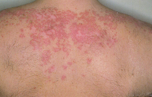
Subacute lupus erythematosus
The eruption corresponds to a profuse form consisting of erythematous and somewhat scaly polycyclic annular plaques which resolve to leave depigmentation and telangiectasia.
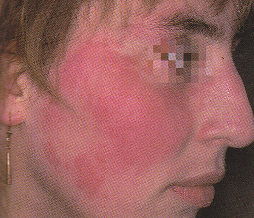
Systemic lupus erythematosus
1. FACE
The eruption is in the form of slightly oedematous erythematous sheets, without atrophy or follicular hyperkeratosis.

2. Fingers
The site of the lesions on the fingers is usually around the nails. The lesions are usually erythematous and telangiectatic, sometimes violet (chilblain-like in appearance).
The lesions are often symmetrical and located on areas exposed to the sun ("butterfly" appearance).
 WhatsApp
 Telegram

 Врачи нашей клиники быстро и абсолютно безболезненно возьмут анализ крови, который будет готов в вашем присутствии.
 Врачи нашей клиники быстро и абсолютно безболезненно возьмут анализ крови на гепатиты В и С.